
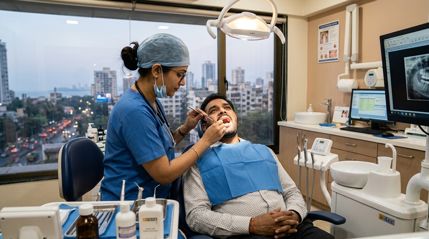
Patients diagnosed with deep pulp inflammation often wonder: where is a root canal tooth located? The short answer is that a root canal tooth can be located anywhere in your upper or lower dental arch. From the most visible front central incisors to the heavy back chewing molars, any tooth in the human mouth contains an internal dental pulp chamber and root canal network. When bacterial decay, trauma, or deep cracks compromise this internal pulp tissue, endodontic therapy saves the natural tooth regardless of its specific anatomical location.
Key Takeaways:
- Universal Tooth Anatomy: All 32 adult teeth possess root canal systems containing nerves and blood vessels, meaning any tooth can require a root canal.
- Front Teeth (Anteriors): Feature single, relatively straight canals; treatments are fast and straightforward, preserving vital smile aesthetics.
- Back Teeth (Molars & Premolars): Possess 2 to 4 intricate, curved canals bearing immense bite force, requiring specialized rotary instrumentation.
- Lifelong Preservation: Saving a tooth at any location preserves alveolar jawbone density and prevents neighboring teeth from tipping and misaligning your bite.
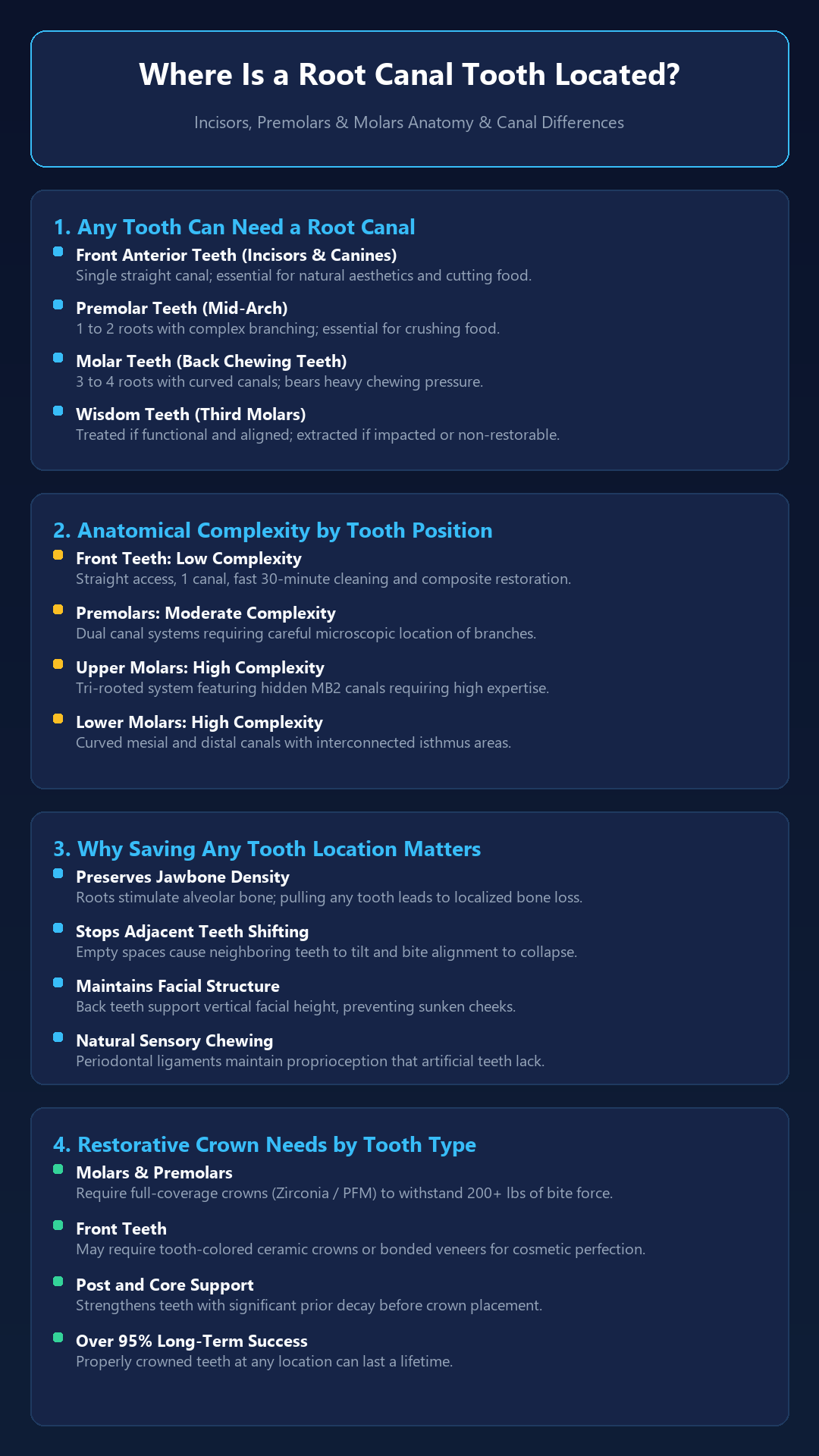
Table of Contents
- Tooth Locations Across the Upper and Lower Dental Arches
- Root Canal Anatomy Differences: Incisors vs. Premolars vs. Molars
- Comparison: Root Canal Complexity by Tooth Location
- Why Preserving a Natural Tooth at Any Location Is Vital
- Restoration and Crown Requirements by Tooth Position
- Managing Complicated or Persistent Root Canal Infections
- Precision Root Canal Therapy in Thane with Dr. Saurabh Pakhale
- Frequently Asked Questions (FAQs)
Tooth Locations Across the Upper and Lower Dental Arches
In a healthy adult dentition with up to 32 teeth, your dentition is divided into four quadrants across the upper (maxillary) and lower (mandibular) arches. Each quadrant features four distinct tooth groups, all of which can undergo root canal therapy:
- Central & Lateral Incisors (Front Smile Teeth): Located right at the front of your smile, these chisel-shaped teeth cut food. They contain single root canals and are easily accessible.
- Canines / Cuspids (Corner Teeth): Located at the corners of your dental arch, having the longest and strongest roots in the entire mouth to guide your jaw motion.
- Premolars / Bicuspids (Mid-Arch Teeth): Positioned between canines and molars, these teeth transition from cutting to crushing food and usually contain one or two distinct canals.
- Molars (Back Chewing Teeth): Located at the far back of the mouth, these large multi-rooted teeth crush and grind food, absorbing up to 200 pounds of force per square inch.
Root Canal Anatomy Differences: Incisors vs. Premolars vs. Molars
Where your affected tooth is located inside the jaw directly influences how endodontic therapy is planned and executed:
1. Anterior Teeth (Incisors and Canines)
Front teeth generally possess one single, wide, and relatively straight root canal. Because access is direct and visible, endodontic cleaning can often be accomplished quickly in 30 to 40 minutes. Aesthetic preservation of the tooth’s natural color is a key priority here.
2. Premolars
Maxillary (upper) first premolars typically have two separate roots or two parallel canals inside a single root. Mandibular (lower) premolars often have one canal that can suddenly bifurcate deep in the apical third, requiring micro-endodontic inspection.
3. Molars (The Workhorses of Chewing)
Upper molars usually have three separate roots and 3 to 4 canals, prominently featuring a hidden fourth canal called the MB2 (mesiobuccal 2). Lower molars typically have two broad roots housing 3 to 4 distinct canals. Molar root canals require specialist expertise because canals can be curved, ribbon-shaped, or interconnected with narrow isthmuses.
Comparison: Root Canal Complexity by Tooth Location
Here is an anatomical breakdown of how root canal procedures differ across various tooth positions:
| Tooth Group & Location | Number of Roots | Typical Canal Count | Anatomical Complexity | Post-RCT Crown Need |
|---|---|---|---|---|
| Front Incisors | 1 Root | 1 Canal | Low (Straight & Wide) | Composite / Aesthetic Ceramic Crown |
| Canines (Cuspids) | 1 Long Root | 1 Canal | Low to Moderate | Composite Restoration or Full Crown |
| Upper Premolars | 1 to 2 Roots | 2 Canals | Moderate (Branching canals) | Full Coverage Crown Essential |
| Lower Premolars | 1 Root | 1 to 2 Canals | Moderate (Deep bifurcations) | Full Coverage Crown Essential |
| Upper & Lower Molars | 2 to 3 Roots | 3 to 4 Canals (MB2) | High (Curved, calcified, micro-channels) | Mandatory Zirconia or PFM Crown |
Why Preserving a Natural Tooth at Any Location Is Vital
No artificial prosthetic—whether a dental implant or bridge—matches the biological function of your natural tooth. Here is why saving a root canal tooth anywhere in your arch is critical:
- Natural Sensory Feedback: The periodontal ligament fibers anchoring natural roots convey tactile feedback to your brain, enabling you to gauge bite pressure when eating.
- Stops Alveolar Bone Shrinkage: Tooth roots continuously stimulate jawbone regeneration. Extracting a tooth causes the underlying jawbone to resorb and thin over time.
- Prevents Unwanted Tooth Drift: Leaving an empty gap allows adjacent teeth to tilt and the opposing tooth to super-erupt, ruining your natural bite.
- Preserves Facial Harmony: Posterior teeth maintain your lower facial vertical dimension, preventing premature sunken cheeks and facial collapse.
Restoration and Crown Requirements by Tooth Position
Once endodontic cleaning and 3D hermetic sealing are complete, restoring structural integrity depends on the tooth’s arch position:
- Back Chewing Teeth (Molars and Premolars): These teeth experience high masticatory shear forces. After a root canal, they require full-coverage dental crowns—such as monolithic Zirconia, E-max ceramic, or Porcelain-Fused-to-Metal (PFM)—to prevent coronal fractures.
- Front Teeth (Incisors): If a front tooth retains substantial healthy enamel with minimal prior decay, a durable direct composite resin bonding may suffice. If severely undermined, an aesthetic all-ceramic crown restores both strength and natural translucency.
Managing Complicated or Persistent Root Canal Infections
If an anatomical anomaly like a calcified branch or hidden lateral canal caused an old treatment to flare up, specialized root canal retreatment can dismantle older fillings, sanitize complex channel networks, and restore complete health to the tooth.
Precision Root Canal Therapy in Thane with Dr. Saurabh Pakhale
Regardless of where your painful tooth is located in your dental arch, receiving gentle, expert endodontic care ensures outstanding clinical results. At Elite Dermadent, Dr. Saurabh Pakhale brings advanced endodontic skill and painless treatment methods to every case.
Using digital radiovisiography, electronic apex measurement, and precision rotary motors, our clinic delivers smooth, stress-free root canal treatment in Thane for incisors, premolars, and complex molars alike.
Frequently Asked Questions (FAQs)
1. Can a front tooth have a root canal?
Yes. Front teeth frequently undergo root canal treatment following sports trauma, direct accidents, or interproximal decay. Because they typically have a single, straight canal, front tooth root canals are fast and have an exceptionally high success rate.
2. Are molar root canals harder than front tooth root canals?
Yes. Molars have multiple curved roots and 3 to 4 narrow canals, including hidden accessory canals like the MB2. They require specialized rotary endodontic equipment and take slightly longer to clean and seal than single-rooted front teeth.
3. Do wisdom teeth ever receive root canal treatment?
Occasionally, yes. If a wisdom tooth (third molar) is fully erupted, functional in biting, properly aligned, and essential for chewing support, a root canal can be performed. However, if it is impacted, misaligned, or difficult to keep clean, extraction is often recommended instead.
4. Does every root canal tooth need a dental crown?
Back teeth (premolars and molars) definitely need a crown because chewing forces will fracture an unprotected, brittle tooth. Front teeth may only need a tooth-colored composite filling if substantial healthy tooth structure remains.
5. Can you tell which tooth has had a root canal just by looking in the mouth?
No. When properly restored with modern composite bonding or high-grade ceramic crowns matching your natural enamel, a root canal tooth looks and feels completely indistinguishable from healthy natural teeth.
No responses yet