

While almost everyone has heard of the procedure, many people often ask: where is the root canal located? Is the “root canal” a surgery, or is it an actual anatomical part of the human tooth? In dental anatomy, the root canal is a natural microscopic channel situated deep within the roots of each tooth. It originates from the central pulp chamber in the tooth’s crown and extends down through the jawbone to the tip of each root. When this inner passage becomes infected by deep cavities or fractures, endodontic therapy cleans and seals this space to permanently save your natural tooth.
Key Takeaways:
- Natural Anatomy, Not Just a Procedure: A root canal is the hollow anatomical pathway inside the root of a tooth that houses dental pulp, blood vessels, and nerve fibers.
- Position in the Jaw: The root canal resides deep below the gum line, securely anchored in the jawbone by the periodontal ligament.
- Canal Variations by Tooth Type: Front teeth typically feature 1 straight root canal, while back molars possess 3 to 4 curved, branching micro-canals.
- Treatment Function: Root canal treatment removes bacterial decay from inside this channel, sanitizes it, and hermetically seals it with biocompatible gutta-percha.
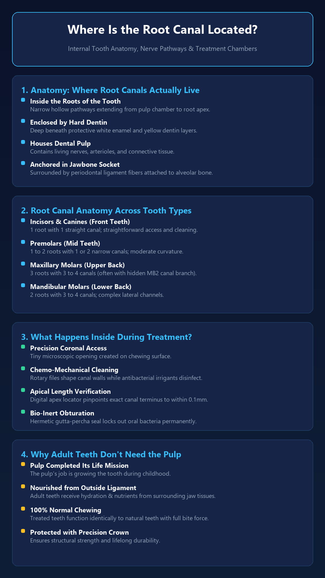
Table of Contents
- Anatomical Location: Where Root Canals Exist Inside the Tooth
- The 4 Layers of Tooth Anatomy: Enamel to the Apical Foramen
- Root Canal Numbers Across Different Teeth in Your Mouth
- What Happens Inside the Root Canal During Treatment?
- Why an Adult Tooth Can Function Perfectly Without Its Pulp
- Complex Canal Anatomy and Endodontic Retreatment
- Microscopic Precision Endodontics with Dr. Saurabh Pakhale
- Frequently Asked Questions (FAQs)
Anatomical Location: Where Root Canals Exist Inside the Tooth
To pinpoint exactly where a root canal is, visualize the two primary sections of a tooth:
- The Crown: The visible portion of the tooth located above the gum line, protected by hard white enamel.
- The Root: The lower portion embedded beneath the gums, anchored into the alveolar socket of the jawbone. Roots comprise approximately two-thirds of the total tooth length.
Deep inside the crown sits the pulp chamber. Extending downwards from this chamber through the center of each root are narrow, microscopic channels known as root canals. At the very tip of each root is a microscopic aperture called the apical foramen, where nerves and tiny blood vessels enter from the jawbone to nourish the tooth during its growth phase.
The 4 Layers of Tooth Anatomy: Enamel to the Apical Foramen
Understanding the exact location of the root canal requires a look through the concentric structural layers of a human tooth:
| Structural Layer | Anatomical Position | Function & Clinical Significance |
|---|---|---|
| 1. Enamel | Outermost layer of the crown above gums | Hardest mineral in the body; shields the tooth from chewing wear and oral acids. |
| 2. Dentin | Beneath enamel in crown and root trunk | Micro-tubular yellowish tissue; transmits hot and cold sensations toward the pulp. |
| 3. Dental Pulp | Central hollow pulp chamber & root canals | Soft living tissue with sensory nerves and blood vessels; target of root canal therapy. |
| 4. Periodontal Ligament | Surrounds outer root surface in jawbone socket | Shock-absorbing connective fibers anchoring root to bone; senses chewing pressure. |
Root Canal Numbers Across Different Teeth in Your Mouth
The complexity and number of root canals vary significantly depending on where the tooth is located in your dental arch:
- Anterior Teeth (Incisors and Canines): Located in the front smile zone. Usually have 1 root and 1 straight canal, making treatment rapid and straightforward.
- Premolars (Bicuspids): Located between canines and molars. Usually possess 1 or 2 roots with 1 to 2 narrow canals.
- Maxillary Molars (Upper Back Chewing Teeth): Large teeth with 3 roots and typically 3 to 4 canals. Upper first molars frequently feature a hidden accessory canal known as the Mesiobuccal 2 (MB2).
- Mandibular Molars (Lower Back Chewing Teeth): Feature 2 roots and 3 to 4 interconnected canals, often exhibiting curved pathways that require flexible instruments.
What Happens Inside the Root Canal During Treatment?
When bacterial decay or trauma breaches the pulp, an endodontist performs root canal treatment directly inside these channels:
- Coronal Access: A tiny opening is gently created on the chewing surface under local anesthesia.
- Debridement & Shaping: Ultra-flexible rotary nickel-titanium (NiTi) files clean out necrotic tissue while warm antimicrobial irrigants flush bacteria from lateral micro-branches.
- Electronic Apex Location: Digital sensors measure canal depth to within 0.1 mm, ensuring the cleaning instruments reach the exact tip of the root without disturbing socket bone.
- 3D Hermetic Sealing: The hollow root space is sealed with biocompatible gutta-percha and bioceramic cement, preventing future bacterial contamination.
Why an Adult Tooth Can Function Perfectly Without Its Pulp
Patients often ask if a tooth is “dead” without its pulp. While the tooth is devitalized (meaning it no longer feels hot or cold), it is not dead in the biological sense. The tooth root remains firmly anchored in the jawbone and continues to receive hydration, blood supply, and mechanical support from the surrounding periodontal ligament and alveolar bone.
Complex Canal Anatomy and Endodontic Retreatment
Due to the complex, branching nature of root canals, a small percentage of cases develop delayed infections if an accessory branch is missed. In such situations, specialized root canal retreatment can reopen the tooth, dissolve old filling material, locate previously hidden canals using dental microscopy, and reseal the root system for lifelong tooth preservation.
Microscopic Precision Endodontics with Dr. Saurabh Pakhale
Understanding your dental anatomy is the first step toward stress-free treatment. Under the expert clinical care of Dr. Saurabh Pakhale, Elite Dermadent is a premier center for advanced, painless root canal treatment in Thane.
Combining digital apex locators, rotary endodontic motors, and custom zirconia crowns, our team delivers predictable, long-lasting tooth preservation.
Frequently Asked Questions (FAQs)
1. Where is the root canal located in the body?
The root canal is an anatomical pathway located inside the roots of your teeth, below the gum line and anchored directly into your upper or lower jawbone.
2. Does every tooth have a root canal?
Yes. Every natural tooth has an internal pulp chamber and between one to four root canals extending through the roots into the jawbone.
3. How many canals are in a back molar tooth?
Back molars typically have three or four root canals spread across two or three roots. Some complex upper molars feature hidden accessory canals like the MB2 canal branch.
4. Can you see a root canal on a dental X-ray?
Yes. On a dental radiograph, root canals appear as thin dark lines running down through the center of the white roots of the tooth. Digital X-rays allow dentists to inspect canal curvature and depth.
5. Why is a crown necessary after a root canal?
Cleaning out the internal pulp and prior decay hollows out the tooth’s core, leaving the outer enamel brittle. A custom dental crown completely covers and protects the remaining tooth structure from cracking under biting forces.
No responses yet